Vascular Interventions and Venous Access
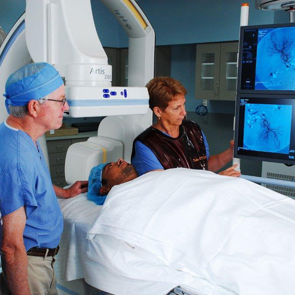
Vascular and Interventional Radiology, a subspecialty of radiology, involves performing minimally invasive procedures with image guidance (e.g., CT, fluoroscopy, ultrasound) to diagnose and treat disease. Interventional Radiology offers patients an alternative to traditional surgery. Most procedures are performed on an outpatient basis, using a local anesthetic or moderate sedation.
The Vascular and Interventional Radiologists at Triad Radiology Associates are fellowship trained and have been awarded certificates of added qualification by the American Board of Radiology. We offer a wide range of vascular and interventional radiology procedures, which include:
• Abdominal Aortic Aneurysms (AAA) and Endoleak Repair
• Peripheral and Visceral Artery Angioplasty and Stenting
• Deep Vein Thrombosis (DVT) and Pulmonary Embolism (PE) Treatments
• IVC Filter Placement and Retrieval
• Insertion and Removal of Implanted Ports, PICC, and Tunneled/Non-Tunneled Catheters
Abdominal Aortic Aneurysms (AAA) and Endoleak Repair
Abdominal aortic aneurysm (AAA) occurs when atherosclerosis or plaque buildup causes the walls of the abdominal aorta to become weak and bulge outward like a balloon. An AAA develops slowly over time and rarely shows symptoms. The larger an aneurysm grows, the more likely it will burst or rupture, causing intense abdominal or back pain, dizziness, nausea or shortness of breath.
To confirm the presence of an AAA, your physician may order imaging tests, such as an abdominal ultrasound, CT, or angiography. Treatment depends on a variety of factors, including the size and location of the aneurysm as well as the patient’s age, kidney function, and overall health.
Certain patients with aneurysms larger than 5 centimeters in diameter can be treated with endovascular abdominal aortic aneurysm repair (EVAR), which is less invasive than surgery and requires placing a stent in the aorta through small incisions in both groins. Following an EVAR, imaging studies are used to closely follow the aneurysm and to evaluate for potential endoleaks. An endoleak is persistent retrograde blood flow into the aneurysm outside the stent from small arterial branches of the aorta. This can sometimes cause the aneurysm to enlarge, potentially leading to rupture. Interventional Radiologists at Triad Radiologists Associates are specially trained to treat endoleaks using minimally invasive techniques.
Sources:
Peripheral and Visceral Artery Angioplasty and Stenting
Angioplasty is a procedure during which a balloon-tipped catheter is used to open a narrowed or blocked blood vessel. Imaging guidance is used to position the catheter across the area of narrowing or blockage within the blood vessel and the balloon is inflated to open the narrowing and improve blood flow. Sometimes, vascular stents, or metal scaffolds, may be needed to help keep the area open. Angioplasty, with or without vascular stenting, is a minimally invasive procedure that usually does not require general anesthesia and can be used to treat both peripheral and visceral artery disease.
Arteries become narrowed overtime as a result of atherosclerosis or plaque, usually in patient who smoke, have diabetes, or high cholesterol. When the arteries of the arms or legs are involved, it is known as Peripheral Artery Disease (PAD) and can result in pain with activity (walking and arm movement), non-healing wounds, numbness, and weakness. When the arteries of the kidney or bowel are involved, it is known as Visceral Artery Disease and can result in hypertension, poor kidney function, and progressive abdominal pain most notable after eating.
Sources:
Deep Vein Thrombosis (DVT) and Pulmonary Embolism (PE) Treatments
Blood clots are semi-solid masses of blood. Normally, blood flows freely through veins and a certain amount of clotting, or coagulation, is necessary and normal. However, when too much clotting occurs, it can lead to serious complications.
When a blood clot forms, it can be stationary (thrombosis) and block blood flow or break loose (embolism) and travel to various parts of the body. Clots that occur in larger veins are called deep vein thrombosis (DVT). A pulmonary embolism (PE) occurs when a major blood vessel in the lung suddenly becomes blocked by a blot clot.
Most times, blood thinning medication is given to treat DVTs and PEs. However, there are times when a large amount of clot causes significant symptoms or can be potentially life threatening. Interventional Radiologists at Triad Radiology Associates are specially trained to treated large clot burdens with minimally invasive techniques. Through small catheters, clot busting medication can be directly introduced into the clot (catheter-directed thrombolysis) or attempts can be made to suction out clot (catheter-based thrombectomy). The procedures help with removing as much clot as soon as possible and require admission in the hospital.
Sources:
IVC Filter Placement and Retrieval
Blood clots in the veins of the legs and pelvis (DVT) can occasionally travel to the lungs where they may cause a pulmonary embolism (PE) or blockage. Bloods clots are usually treated with blood thinning medication. However, in patients who are bleeding and cannot receive blood thinning medication, who have large clot burdens, or who develop more clots while taking blood thinning medication, Inferior Vena Cava (IVC) filters can be placed, which help to reduce the risk of pulmonary embolism by trapping large clots and preventing them from reaching the heart and lungs, which can sometimes be life-threatening.
An IVC filter is a small umbrella shaped device that is placed in the large vein within the abdomen that drain the legs. It is placed through a small catheter inserted from a small nick in the neck or groin. Most IVC filters can be temporarily placed and retrieved while others may be permanent. When the filter is no longer needed, the retrieval IVC filters can usually be removed using the same minimally invasive techniques used during placement.
Sources:
A venous access procedure (also called vascular access procedure) is used for patients who have conditions that require frequent intravenous (IV) treatments. The procedure involves the insertion of a flexible and sterile thin plastic tube, or catheter, into a blood vessel to provide an effective method of drawing blood or delivering medications, blood products, or nutrition into a patient’s bloodstream over various periods of time (e.g., weeks, months, years).
The major types of venous access catheters include
- Peripherally inserted central catheter (PICC): a long catheter that extends from an arm or leg vein into the largest vein (superior vena cava or inferior vena cava) near the heart. Typically, it provides central IV access for several weeks, however, it may remain in place for several months.
- Tunneled catheter: usually has a cuff that stimulates tissue growth that will help hold it in place in the body. These catheters come in various sizes and are secure and easy to access. Both the tunnel and the cuff help decrease the risk of catheter infection.
- Non-tunneled catheter: is often larger in caliber than a PICC and is designed to be placed via a relatively large, more central vein (e.g., jugular vein, femoral vein).
- Subcutaneous implantable port: consists of a catheter attached to a small reservoir, both of which are placed under the skin similar to tunneled catheters.
Other equipment that may be used during the procedure includes an intravenous line (IV), ultrasound machine, and devices that monitor your heartbeat and blood pressure.
Sources:
Triad Radiology offers Vascular Interventions and Venous Access at a variety of locations, including hospitals, imaging centers, and clinics. Contact us if you want to learn more or schedule an appointment.


